

GIST Pathology Samples and Processing
When Is a Biopsy Done Before Surgery for GIST?
Sometimes GIST diagnosis is made using biopsy samples obtained before any surgery is attempted. This is done when knowing the diagnosis will change the next step in treatment. For example, if a small tumor might be a type that would not require removal, then the biopsy results could allow unnecessary surgery to be avoided. For large tumors, if pre-surgery treatment with imatinib might shrink the tumor and reduce the scope of surgery, or if shrinkage might make it possible to resect a tumor that currently appears inoperable, then biopsy is definitely needed to diagnose GIST before imatinib can be prescribed.
Endoscopic biopsies
An endoscope is a flexible fiberoptic viewing device that can be passed through the mouth into the stomach or duodenum, or through the anus into the rectum or colon. (The endoscope cannot reach most parts of the small intestine.) An ultrasound device in the endoscope is used to image the tumor behind the lining of the GI tract to determine where to sample it. Because these procedures are done through the GI tract, there is no risk of releasing tumor cells into the abdominal cavity.
Endoscopic ultrasound fine needle aspiration (EUS-FNA): A very thin needle attached to a syringe is inserted from inside the digestive tract through the GI tract wall and into the tumor. The doctor pulls back on the syringe plunger to obtain a sample of the tumor. The small amounts of tissue obtained are sent to the pathologist for examination and analysis.
Forceps biopsy: For the rare GISTs that protrude into the interior of the digestive organ, a forceps inserted through the endoscope can pinch off a biopsy sample to send to the pathologist.
Core needle biopsy
A larger needle is inserted through the skin of the abdomen into the tumor, using CT to help guide the needle to the correct location. The core needle is a double needle with a hollow cutting needle inside the outer needle. The interior cutting tube punches a sample from the tumor, and this core is removed through the outer needle. This procedure yields a larger sample than fine-needle aspiration. Core needle biopsy provides more tissue for analysis, which can aid in the diagnosis.
When Is a GIST Diagnosis Made on a Surgically Resected Tumor Rather Than on a Biopsy?
If a pre-surgery biopsy would not change the surgeon’s plans to remove (resect) the tumor, then the diagnosis may be postponed until after the operation. For example, this might happen when a tumor is large enough to require removal regardless of what type it is, and pretreatment with imatinib is not needed to improve resectability.
All the tissue removed during an operation is called the surgical specimen. The entire specimen is sent to the surgical pathologist for examination. The pathologist selects areas of the specimen to examine under the microscope.
How Are Pathology Specimens Prepared for Analysis?
When a biopsy sample or a surgical specimen arrives at the pathology department, the pathologist follows this general sequence of steps.
In the case of a biopsy, the entire specimen is used for analysis. In the case of a surgical resection specimen, sampling occurs so that representative portions of the tumor can be analyzed. The portions of tissue are cut very thin and placed in a plastic cassette. These cassettes are fixed in formalin which makes the tissue more firm. The specimens are then placed on a machine that helps further fix the tissue with heat and pressure and then uses a series of solvents to treat the tissue and remove lipids and allow wax (called paraffin) to be suffused into the tissue. This is done overnight and then early the next morning the tissue is embedded in a larger block of wax that will allow the tissue to be cut very thin on a special instrument called a microtome and then placed on a glass slide. These slides are then de-paraffinized so that the tissue can be stained with water-based dyes and other reagents. The initial stain is with hematoxylin and eosin (H&E) that stains cell nuclei (containing the DNA) blue and stains the cytoplasm and most other structures pink. These stains allow visualization of the shapes of the cells and the architecture of the tissue, which helps the pathologist to make a diagnosis. These stains also allow the pathologist to determine the mitotic rate, which suggests how fast the tumor is growing. In the case of GIST, additional immunohistochemical tests are then performed to look for expression of KIT and other protein markers, to help confirm the diagnosis.
Making Patient Slides
The processing of tumor tissue into slides is illustrated in these photos.
| The tumor is sectioned so that tumor and margins can be sampled properly. | 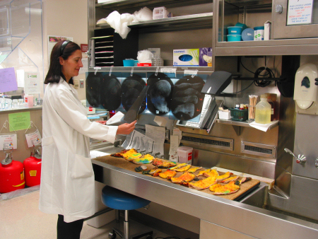 |
| Small sections of tissue about the size of a nickel (about 2 cm) are placed in labeled plastic cassettes and fixed in formalin. | 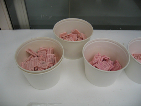 |
| The cassettes are further fixed and suffused with paraffin wax in an automatic process. | 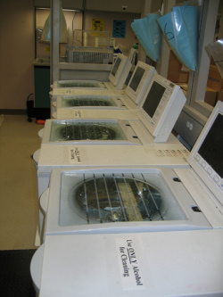 |
| The treated tissue is then oriented and embedded in paraffin in a mold. | 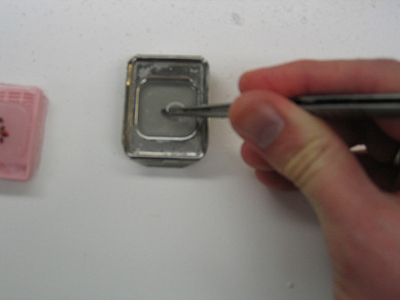 |
| Next it is cooled to adhere to the labeled tissue cassette. | 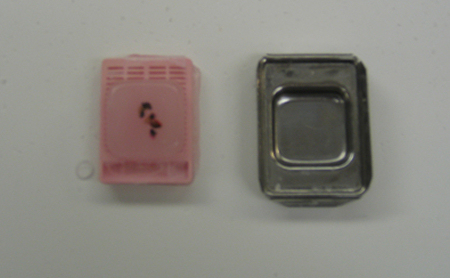 |
| These blocks are then cut very thin on a microtome. | 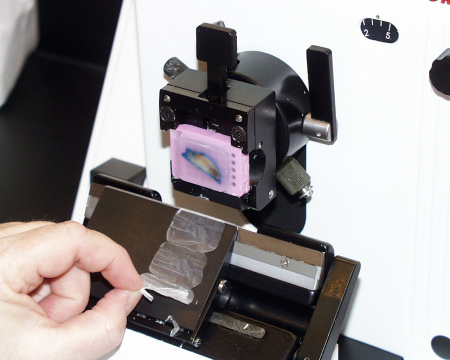 |
| Then the ribbon of sections is floated in a water bath and lifted onto a labeled glass slide. | 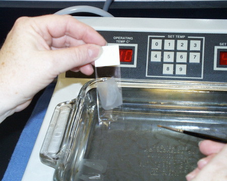 |
| These slides are then stained with hematoxylin and eosin (H&E). | 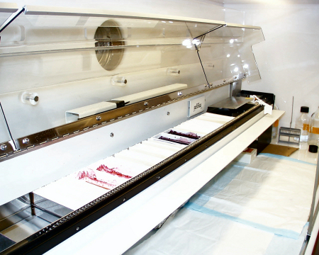 |
| Stained slides are coverslipped using an automated process. | 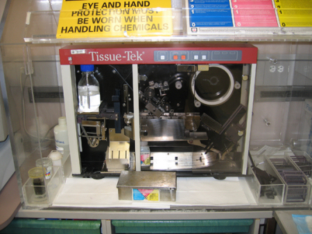 |
| Now the stained slides are ready. | 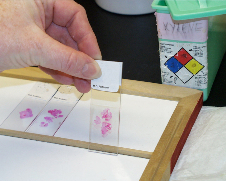 |
| Finally the slides are delivered to the pathologist for diagnosis. | 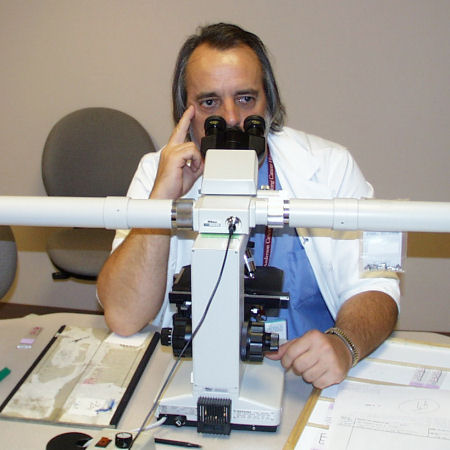 |
If additional immunohistochemical studies are needed, further sli
des can be cut for this purpose. After final diagnosis, the glass slides and paraffin blocks are carefully stored for future reference or additional studies. The extra stored material is also extremely important for future research.
How long do these sample preparations take?
For small biopsy samples, tissue processing, special studies, and interpretation usually takes between 4 and 6 days. For surgical resection specimens, this entire process takes somewhat longer, usually greater than a week or so.
How quickly will my pathology results be available?
As we discussed, the technical portion of the analysis can take up to a week. The pathologist then sends a report your physician, usually a surgeon and/or medical oncologist. They may need to discuss the case at an internal conference to produce a full interpretation. In a small number of cases, materials may need to be sent out for additional testing or review by an expert pathologist, and this will extend the time to a final diagnosis. If molecular analysis of the KIT or PDGFRA gene is needed, this can often take two weeks or more after the initial diagnosis. These genetic tests are only performed at a small number of specialized centers.
Go to next section Pathology Analyses for GIST